Active fraction (HS7) from Taiwanofungus camphoratus inhibits AKT-mTOR, ERK and STAT3 pathways and induces CDK inhibitors in CL1-0 human lung cancer cells
Abstract
Figures 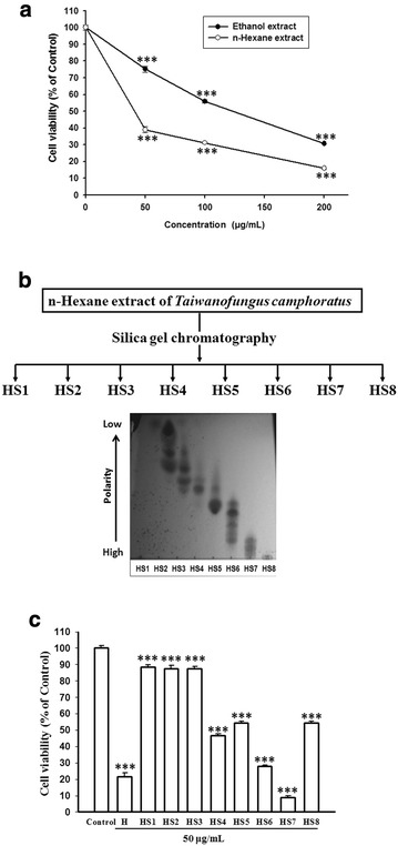
Fig. 1
The activities of ethanol and…
Fig. 1
The activities of ethanol and n -hexane extracts of T. camphoratus and the…
Fig. 1 The activities of ethanol and n-hexane extracts of T. camphoratus and the eight separated fractions from n-hexane extract on the growth inhibition of CL1-0 lung cancer cells. a The cell viabilities of CL1-0 cells were measured after treatment with the ethanol or n-hexane extract of T. camphoratus for 72 h. b Eight fractions (HS1–HS8) were separated from the n-hexane extract by silica gel chromatography according to their polarities displayed in thin-layer chromatography. c The cell viabilities of CL1-0 cells were measured after treatment with n-hexane extract (H) or the separated fraction (HS1–HS8) at dose of 50 μg/mL as indicated for 72 h. Cell viability data (mean ± SE) are expressed as a percentage compared to the control and analyzed as described in “Statistical analysis”. ***p < 0.001 compared to the control group
Fig. 2
The effects of gefitinib (Iressa)…
Fig. 2
The effects of gefitinib (Iressa) and HS7 on the proliferation of CL1-0 lung…
Fig. 2 The effects of gefitinib (Iressa) and HS7 on the proliferation of CL1-0 lung cancer cells. a The cell viability of CL1-0 cells was measured after treatment with gefitinib for 72 h. b The cell viability of CL1-0 cells was measured after treatment with HS7 for 72 h. c The effect of HS7 on CL1-0 cells was examined by phase-contrast microscopy after 72 h of treatment, scale bar = 100 μm. Cell viability data (mean ± SE) are expressed as a percentage compared to the control and analyzed as described in “Statistical analysis”. ***p < 0.001 compared to the control group
Fig. 3
The effect of HS7 on…
Fig. 3
The effect of HS7 on the cell viability of MRC-5 normal fetal human…
Fig. 3 The effect of HS7 on the cell viability of MRC-5 normal fetal human lung fibroblasts. a The cell viability of MRC-5 cells was measured after treatment with HS7 for 72 h. b The effect of HS7 on MRC-5 cells was examined by phase-contrast microscopy after 72 h of treatment, scale bar = 100 μm. Cell viability data (mean ± SE) are expressed as a percentage compared to the control and analyzed as described in “Statistical analysis”
Fig. 4
Representative flow cytometry histograms of…
Fig. 4
Representative flow cytometry histograms of CL1-0 lung cancer cells after treatment with HS7…
Fig. 4 Representative flow cytometry histograms of CL1-0 lung cancer cells after treatment with HS7 for 72 h. HS7 increased the apoptotic sub-G1 fraction in CL1-0 cells at dose of 25 μg/mL but did not significant change the distribution of cells in the G0/G1, S and G2/M phases of the cell cycle within this dose. The percentage of cells in different phases of the cell cycle is shown in Table 1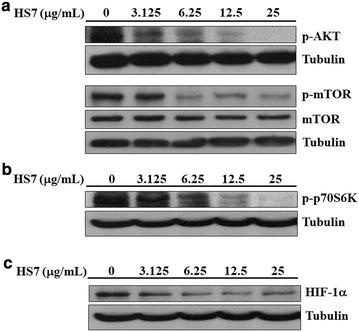
Fig. 5
Inhibitory effects of HS7 on…
Fig. 5
Inhibitory effects of HS7 on the AKT-mTOR signaling pathway and its downstream effectors…
Fig. 5 Inhibitory effects of HS7 on the AKT-mTOR signaling pathway and its downstream effectors in CL1-0 lung cancer cells after 72 h of treatment. a HS7 decreased the protein levels of phosphorylated AKT (p-AKT) and phosphorylated mTOR (p-mTOR) in a dose-dependent manner. b HS7 dose-dependently decreased the phosphorylated p70S6K protein (p-p70S6K). c HS7 dose-dependently decreased the HIF-1α protein. Cell lysates were analyzed by Western blot, using tubulin as loading control
Fig. 6
Inhibitory effects of HS7 on…
Fig. 6
Inhibitory effects of HS7 on the ERK and STAT3 signaling pathways in CL1-0…
Fig. 6 Inhibitory effects of HS7 on the ERK and STAT3 signaling pathways in CL1-0 lung cancer cells after 72 h of treatment. a HS7 decreased the protein levels of phosphorylated ERK (p-ERK) in a dose-dependent manner. b HS7 dose-dependently decreased both the protein levels of total (STAT3) and phosphorylated STAT3 (p-STAT3). Cell lysates were analyzed by Western blot, using tubulin or GAPDH as loading control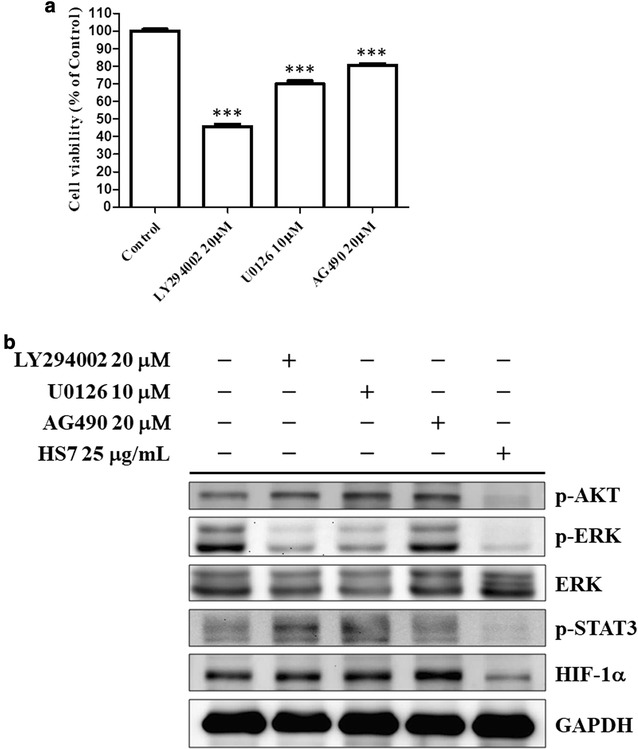
Fig. 7
Effects of synthetic inhibitors and…
Fig. 7
Effects of synthetic inhibitors and HS7 on the AKT ERK and STAT3 signaling…
Fig. 7 Effects of synthetic inhibitors and HS7 on the AKT ERK and STAT3 signaling pathways in CL1-0 lung cancer cells after 72 h of treatment. a The cell viabilities of CL1-0 cells were measured after treatment with LY294002 (PI3K-AKT inhibitor), U0126 (MEK-ERK inhibitor) or AG490 (JAK-STAT3 inhibitor) at indicated doses for 72 h. Cell viability data (mean ± SE) are expressed as a percentage compared to the control and analyzed as described in “Statistical analysis”. ***p < 0.001 compared to the control group. b The effects of LY294002, U0126, AG490 and HS7 on the protein levels of phosphorylated AKT (p-AKT), phosphorylated ERK (p-ERK), ERK, phosphorylated STAT3 (p-STAT3) and HIF-1α in CL1-0 cells. Cell lysates were analyzed by Western blot, using GAPDH as loading control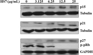
Fig. 8
HS7 induces CDK inhibitors and…
Fig. 8
HS7 induces CDK inhibitors and inhibits phosphorylation of pRb protein in CL1-0 lung…
Fig. 8 HS7 induces CDK inhibitors and inhibits phosphorylation of pRb protein in CL1-0 lung cancer cells after 72 h of treatment. HS7 dose-dependently increased protein levels of p15, p21 and p27, and decreased phosphorylated pRb protein (p-pRb). Cell lysates were analyzed by Western blot, using tubulin or GAPDH as loading control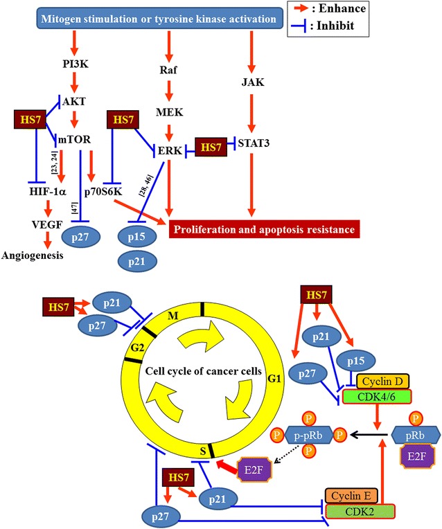
Fig. 9
Schematic diagram displays the proposed…
Fig. 9
Schematic diagram displays the proposed mechanisms of action of HS7 in CL1-0 lung…
Fig. 9 Schematic diagram displays the proposed mechanisms of action of HS7 in CL1-0 lung cancer cells. Through inhibition of AKT-mTOR, ERK and STAT3 signaling pathways, the active fraction HS7 from T. camphoratus increases the CDK inhibitors (p15, p21 and p27) and leads to cell cycle suppression and apoptosis induction. References for the proposed inhibition or enhancement are indicated along the arrow or T-bar. ERK extracellular signal-regulated kinases, HIF-α hypoxia-inducible factor 1-alpha, JAK Janus kinase, MEK mitogen-activated protein/extracellular signal-regulated kinase, mTOR mammalian target of rapamycin, p70S6K ribosomal p70 S6 kinase, PI3K phosphoinositide 3-kinase, Raf rapidly accelerated fibrosarcoma, STAT3 signal transducer and activator of transcription 3, VEGF vascular endothelial growth factor All figures (9)