Anti-Inflammatory Effect of Medicinal Fungus Antrodia cinnamomea Cultivated on Pinus morrisonicola Hayata
Abstract
Figures 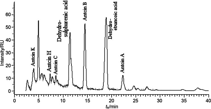
Fig. 1
The chromatogram of triterpenoids found…
Fig. 1
The chromatogram of triterpenoids found in Antrodia cinnamomea. Peaks were detected using UV…
Fig. 1 The chromatogram of triterpenoids found in Antrodia cinnamomea. Peaks were detected using UV detector at λ=254 nm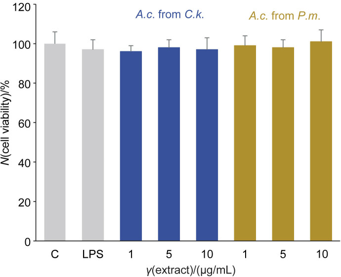
Fig. 2
Cell viability test for the…
Fig. 2
Cell viability test for the Antrodia cinnamomea ( A.c .) extracts. BV-2 cells…
Fig. 2 Cell viability test for the Antrodia cinnamomea (A.c.) extracts. BV-2 cells were treated with γ(extract)=1, 5 or 10 μg/mL for 24 h. The MTT results showed no changes in the cell viability. Values represent the mean±S.D., N=3. C=untreated control, LPS=lipopolysaccharide-stimulated BV-2 cells, C.k.=Cinnamomum kanehirae, P.m.=Pinus morrisonicola Hayata disc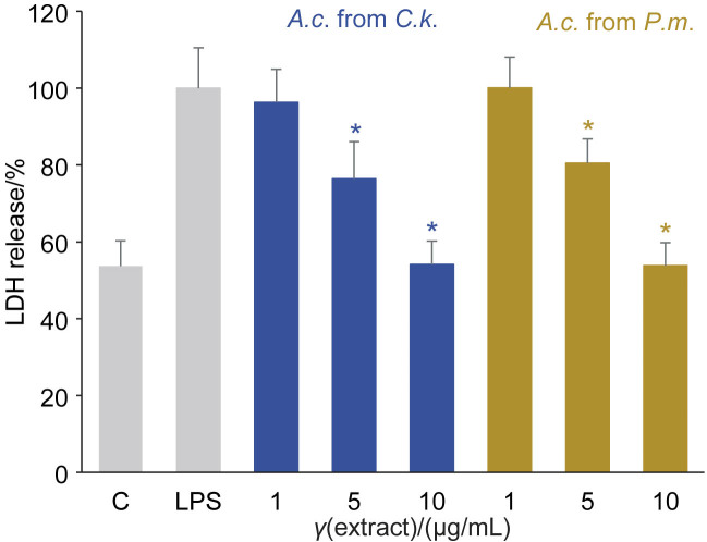
Fig. 3
Protection from cell injury. Cells…
Fig. 3
Protection from cell injury. Cells were treated with lipopolysaccharide γ (LPS)=3 μg/mL alone…
Fig. 3 Protection from cell injury. Cells were treated with lipopolysaccharide γ(LPS)=3 μg/mL alone and/or in combination with Antrodia cinnamomea (A.c.) extracts (γ=1, 5 or 10 μg/mL) for 24 h. The release of lactate dehydrogenase (LDH) expressed as a percentage of LPS as control was reduced significantly by both extracts. Values represent the mean±S.D., N=3. C=untreated control, C.k.=Cinnamomum kanehirae, P.m.=Pinus morrisonicola Hayata. *Significant difference between the LPS alone and LPS with A. cinnamomea extracts determined using Scheffe's test (p<0.05)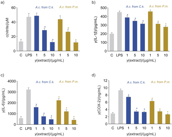
Fig. 4
Comparison of anti-inflammatory activity of…
Fig. 4
Comparison of anti-inflammatory activity of the two types of Antrodia cinnamomea (A.c .)…
Fig. 4 Comparison of anti-inflammatory activity of the two types of Antrodia cinnamomea (A.c.) extracts: a) nitrogen monoxide (NO), b) interleukin (IL)-1β, c) IL-6 and d) cyclooxygenase-2 (COX-2) shows the indicated inflammatory mediators. BV-2 cells were treated with 3 μg/mL lipopolysaccharide (LPS) alone or in combination with the extracts of 1, 5 or 10 μg/mL fruiting body of A. cinnamomea grown on Cinnamomum kanehirae (C.k.) or Pinus morrisonicola Hayata (P.m.) disc for 24 h. Supernatants of the treated and untreated cell cultures were collected and analysed as described in the methods. Results are presented as the mean value±S.D., N=3. C=untreated control. *Significant difference between the LPS alone and LPS with A. cinnamomea extracts determined using Scheffe's test (p<0.05)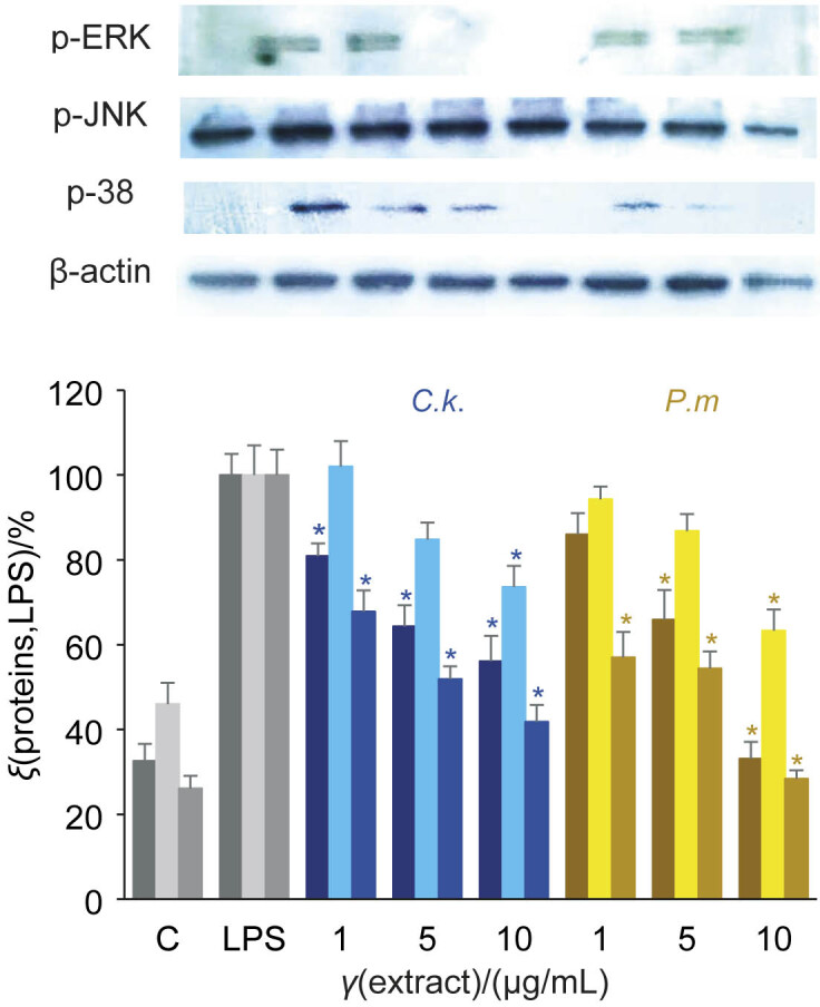
Fig. 5
Suppression of mitogen-activated protein kinase…
Fig. 5
Suppression of mitogen-activated protein kinase (MAPK) signalling pathway. BV-2 cells stimulated with 3…
Fig. 5 Suppression of mitogen-activated protein kinase (MAPK) signalling pathway. BV-2 cells stimulated with 3 μg/mL lipopolysaccharide (LPS) alone or in combination with the extracts of Antrodia cinnamomea γ=1, 5 or 10 μg/mL for 30 min. Cells were extracted and analysed by Western blotting. The results showed that the extracts of the fruiting body of A. cinnamomea grown on Cinnamomum kanehirae (C.k.) or Pinus morrisonicola (P.m.) disc had a similar pattern of suppression of p38, JNK and ERK/MAPK signalling pathways. Results represent the mean value±S.D., N=3. C=untreated control. *Significant difference between the LPS alone and LPS with the extracts of A. cinnamomea determined using Scheffe's test (p<0.05)