2,4-Dimethoxy-6-Methylbenzene-1,3-diol, a Benzenoid From Antrodia cinnamomea, Mitigates Psoriasiform Inflammation by Suppressing MAPK/NF-κB Phosphorylation and GDAP1L1/Drp1 Translocation
Abstract
Antrodia cinnamomea exhibits anti-inflammatory, antioxidant, and immunomodulatory activities. We aimed to explore the antipsoriatic potential of 2,4-dimethoxy-6-methylbenzene-1,3-diol (DMD) derived from A. cinnamomea. The macrophages activated by imiquimod (IMQ) were used as the cell model for examining the anti-inflammatory effect of DMD in vitro. A significantly high inhibition of IL-23 and IL-6 by DMD was observed in THP-1 macrophages and bone marrow-derived mouse macrophages. The conditioned medium of DMD-treated macrophages could reduce neutrophil migration and keratinocyte overproliferation. DMD could downregulate cytokine/chemokine by suppressing the phosphorylation of mitogen-activated protein kinases (MAPKs) and NF-κB. We also observed inhibition of GDAP1L1/Drp1 translocation from the cytoplasm to mitochondria by DMD intervention. Thus, mitochondrial fission could be a novel target for treating psoriatic inflammation. A psoriasiform mouse model treated by IMQ showed reduced scaling, erythema, and skin thickening after topical application of DMD. Compared to the IMQ stimulation only, the active compound decreased epidermal thickness by about 2-fold. DMD diminished the number of infiltrating macrophages and neutrophils and their related cytokine/chemokine production in the lesional skin. Immunostaining of the IMQ-treated skin demonstrated the inhibition of GDAP1LI and phosphorylated Drp1 by DMD. The present study provides insight regarding the potential use of DMD as an effective treatment modality for psoriatic inflammation.
Figures 
Figure 1
DMD inhibit IMQ-induced inflammatory cytokines…
Figure 1
DMD inhibit IMQ-induced inflammatory cytokines in THP-1 and BMDMs. (A, B) Cell viability…
Figure 1 DMD inhibit IMQ-induced inflammatory cytokines in THP-1 and BMDMs. (A, B) Cell viability in THP-1 cells determined by MTT and trypan blue assays after incubation with IMQ and DMD in the indicated concentration, respectively, for 24 h (n=3). (C) RT-qPCR analysis of the cytokines in IMQ-stimulated THP-1 cells following DMD treatment (n=3). (D) ELISA analysis of the cytokines in IMQ-stimulated THP-1 cells following DMD treatment (n=3). (E) ELISA analysis of the cytokines in IMQ-stimulated BMDMs following DMD treatment (n=3). The data shown are representative of three independent experiments. GAPDH is served as an internal control. *P < 0.05, **P < 0.01, and ***P < 0.001 when compared to IMQ group. Data are represented as mean ± SEM.
Figure 2
DMD inhibit IMQ-induced activation in…
Figure 2
DMD inhibit IMQ-induced activation in THP-1 cells through MAPK and NF-κB pathways, and…
Figure 2 DMD inhibit IMQ-induced activation in THP-1 cells through MAPK and NF-κB pathways, and block neutrophil invasion and keratinocyte proliferation. (A) RT-qPCR analysis of the chemokines in IMQ-stimulated THP-1 cells following DMD treatment (n=4). (B) RT-qPCR analysis of GDAP1L1 in IMQ-stimulated THP-1 cells following DMD treatment (n=4). (C–E) THP-1 cells were treated with IMQ in the presence or absence of DMD. Immunoblotting analysis of phosphorylation of NF-κB, MAPKs, and GDAP1L1 was determined, respectively. (F) Neutrophil migration was measured by wound healing assay. Neutrophils were treated with the conditioned medium of THP-1 cells for 24 h to determine the rate of migration into the scratched area. Wound healing was quantified by measuring the distance between scratch edges at 0 and 24 h. (G) Neutrophil invasion was measured by the Boyden chamber invasion assay. Neutrophils were loaded on the top chambers. The cells were allowed to invade for 4 h, and the rate of invasion was quantified (n=4). (H) Keratinocyte proliferation induced by the conditioned medium of THP-1 cells was measured using the MTT and trypan blue assays (n=3). (I) Immunoblotting of the phosphorylation of STAT3 in keratinocytes stimulated with the conditioned medium of THP-1 cells for 24 h. One out of three independent experiments is shown. *P < 0.05, **P < 0.01, and ***P < 0.001 when compared to IMQ group. Data are represented as mean ± SEM.
Figure 3
DMD inhibit IMQ-induced activation in…
Figure 3
DMD inhibit IMQ-induced activation in THP-1 cells through decreasing Drp1 phosphorylation and GDAP1L1/Drp1…
Figure 3 DMD inhibit IMQ-induced activation in THP-1 cells through decreasing Drp1 phosphorylation and GDAP1L1/Drp1 translocation. (A) Immunoblotting of GDAP1L1 and Drp1 S616 in IMQ-stimulated THP-1 cells following DMD treatment. (B) THP-1 cells were pretreated by DMD or TBHQ and then stimulated with IMQ or menadione for 2 h. Cells were fractionated into cytosolic and mitochondrial fractions and subjected to immunoblotting assay to determine GDAP1L1 and Drp1. (C) THP-1 cells were pretreated by DMD or TBHQ and then stimulated with IMQ or menadione for 2 h. Cells were fractionated into cytosolic and mitochondrial fractions and subjected to immunoblotting assay to determine Drp1 and its phosphorylated form (Drp1 S616). (D) THP-1 cells were stimulated with IMQ for 2 h, and then stained with MitoTracker Red. Cells were fixed and immunostained with the antibody against Drp1 S616 followed by confocal microscopy. Average mitochondrial length (μm) and comparison of mitochondrial length distribution in the indicated groups is shown in the right panel. (E) Immunoprecipitation of GDAP1L1 and Drp1 in mitochondria of THP-1 cells as shown in the immunoblotting. (F) THP-1 cells were stimulated with IMQ for 2 h, and then stained with MitoTracker Red. Cells were fixed and immunostained with the antibody against GDAP1L1 or Drp1 S616 followed by confocal microscopy. Scale bars, 10 μm. All experiments were repeated two or three times with similar results.
Figure 4
DMD attenuate IMQ-induced psoriasis-like inflammation…
Figure 4
DMD attenuate IMQ-induced psoriasis-like inflammation in a mouse model. (A) Scheme of the…
Figure 4 DMD attenuate IMQ-induced psoriasis-like inflammation in a mouse model. (A) Scheme of the experimental protocol for AC or DMD treatment in IMQ-induced psoriasis-like inflammation model in mice. (B) Phenotypical and microscopic images of IMQ-induced psoriasis-like inflammation on mouse skin with and without the treatment by AC or DMD after 5 days. (C) The cumulative score (scaling plus erythema from 0 to 4 each) is depicted. (D) TEWL was measured on Day 6. (E) Epidermal thicknesses measured according to H&E-stained histology. (F) Munro’s microabscesses measured using an image analysis system. All experiments were performed at least three times. **P < 0.01 and ***P < 0.001 when compared to IMQ group. Scale bar, 100 μm. Data are represented as mean ± SEM. (n=6).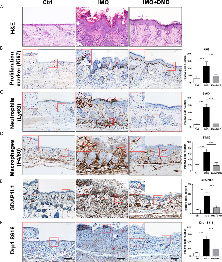
Figure 5
DMD attenuate IMQ-induced psoriasis-like inflammation…
Figure 5
DMD attenuate IMQ-induced psoriasis-like inflammation in a mouse model as observed by skin…
Figure 5 DMD attenuate IMQ-induced psoriasis-like inflammation in a mouse model as observed by skin histology. (A) Histological assessment of the skin by H&E staining. (B) Histological assessment of the skin by Ki67 staining. (C) Histological assessment of the skin by Ly6G staining. (D) Histological assessment of the skin by F4/80 staining. (E) Histological assessment of the skin by GDAP1L1 staining. (F) Histological assessment of the skin by Drp1 S616 staining. The upper left corner of each image indicates the higher power magnification of the red frame. The red arrow indicates the location of the antibody-stained cells. The quantification of the antibody-positive cells is shown in the right panel of each figure. ***P < 0.001 when compared to IMQ group. Data are represented as mean ± SEM. (n=9).
Figure 6
DMD suppress the expression of…
Figure 6
DMD suppress the expression of proinflammatory mediators in IMQ-induced psoriasiform lesion in mice.…
Figure 6 DMD suppress the expression of proinflammatory mediators in IMQ-induced psoriasiform lesion in mice. (A–G) The dorsal skin of mouse was topically treated with IMQ cream for 5 consecutive days. AC or DMD was also topically applied on the lesional skin if necessary. Total skin RNA was extracted and the mRNA level of IL-23, IL-6, IL-17A, IL-24, TNF, CXCL2, and F4/80 by RT-qPCR (n=8−12), respectively. *P < 0.05, **P < 0.01, ***P< 0.001 when compared to IMQ group. Data are represented as mean ± SEM. (n=6).
Figure 7
Schematic representation of the proposed…
Figure 7
Schematic representation of the proposed mechanisms responsible for the anti-inflammatory effect of DMD…
Figure 7 Schematic representation of the proposed mechanisms responsible for the anti-inflammatory effect of DMD in macrophages. We found that IMQ stimulation induced GDAP1L1 expression and GDAP1L1-dependent Drp1 S616 phosphorylation, and then caused mitochondria translocation of GDAP1L1 and phosphorylated Drp1 S616, leading to mitochondrial fission. IMQ stimulation also induced inflammatory mediators’ expression as a result of the activation of NF-κB and MAPK phosphorylation, which was the GDAP1L1-dependent manner. We demonstrate that DMD prevented IMQ-induced mitochondrial fission by blocking GDAP1L1-dependent Drp1 S616 phosphorylation and mitochondria translocation. It could suppress proinflammatory mediator expression in activated macrophages. DMD treatment suppressed migration of activated macrophages into the inflamed site of the mouse skin. The cytokines and chemokines from macrophages in turn rescue neutrophils and cause epidermal proliferation were blocked by DMD treatment. All figures (7)