CCM111 prevents hepatic fibrosis via cooperative inhibition of TGF-β, Wnt and STAT3 signaling pathways
Abstract
CCM111 is an aqueous extract of Antrodia cinnamomea (AC) that has exhibited anti-liver fibrosis functions. However, the detailed mechanisms of AC action against liver fibrosis have not been elucidated yet. The present research showed that CCM111 significantly lowered the levels of the hepatic enzyme markers glutamate oxaloacetate transaminase (GOT) and glutamic pyruvic transaminase (GPT), prevented liver damage and collagen deposition, and downregulated TGF-β/Smad signaling in a dose-dependent manner compared with CCl4 treatment alone. CCM111 markedly inhibited TGF-β, Wnt and STAT3 signaling pathway-regulated downstream genes in the liver by next-generation sequencing. The antifibrotic mechanisms of CCM111 were further demonstrated in HSC-T6 cells. Our data demonstrated for the first time that CCM111 can protect against CCl4-induced liver fibrosis by the cooperative inhibition of TGF-β-, Wnt- and STAT3-dependent proinflammatory and profibrotic mediators, suggesting that CCM111 might be a candidate for preventing and treating chronic fibrotic liver diseases.
Figures 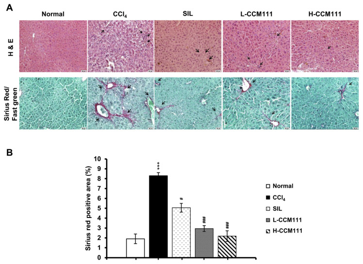
Fig. 1
CCM111 suppressed the liver morphology…
Fig. 1
CCM111 suppressed the liver morphology in CCl 4 -treated animals. (A) Images of…
Fig. 1 CCM111 suppressed the liver morphology in CCl4-treated animals. (A) Images of liver tissue sections stained separately with H&E stain and Sirius Red/Fast Green stain. The sections were imaged at 200× magnification. (B) Quantification of Sirius Red/Fast Green staining. All values are expressed as the mean ± SD (n = 6). Student’s t-test: *P-value < 0.05, **P-value < 0.01, ***P-value < 0.001 for Normal vs. CCl4. #P-value < 0.05, ##P-value < 0.01, ###P-value < 0.001, for CCl4 vs. SIL, L-CCM111, or H-CCM111, respectively.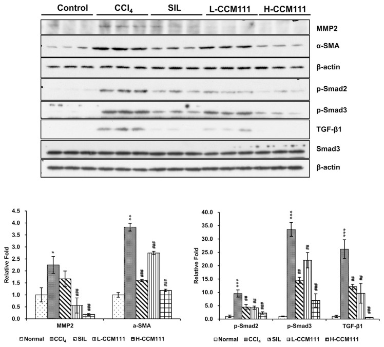
Fig. 2
CCM111 reduced TGF-β1-induced expression of…
Fig. 2
CCM111 reduced TGF-β1-induced expression of α-SMA and MMP2 and activation of the TGF-β…
Fig. 2 CCM111 reduced TGF-β1-induced expression of α-SMA and MMP2 and activation of the TGF-β signaling pathway in CCl4-treated animals. Total liver cell lysates were analyzed for α-SMA, MMP2, p-Smad2, p-Smad3, TGF-β1, and Smad3 protein expression by western blot. β-Actin was the loading control. The quantification data are reported as the mean ± standard deviation of three independent experiments. Student’s t-test: *P-value < 0.05, **P-value < 0.01, ***P-value < 0.001 for Normal vs. CCl4. #P-value < 0.05, ##P-value < 0.01, ###P-value < 0.001, for CCl4 vs. SIL, L-CCM111, or H-CCM111, respectively.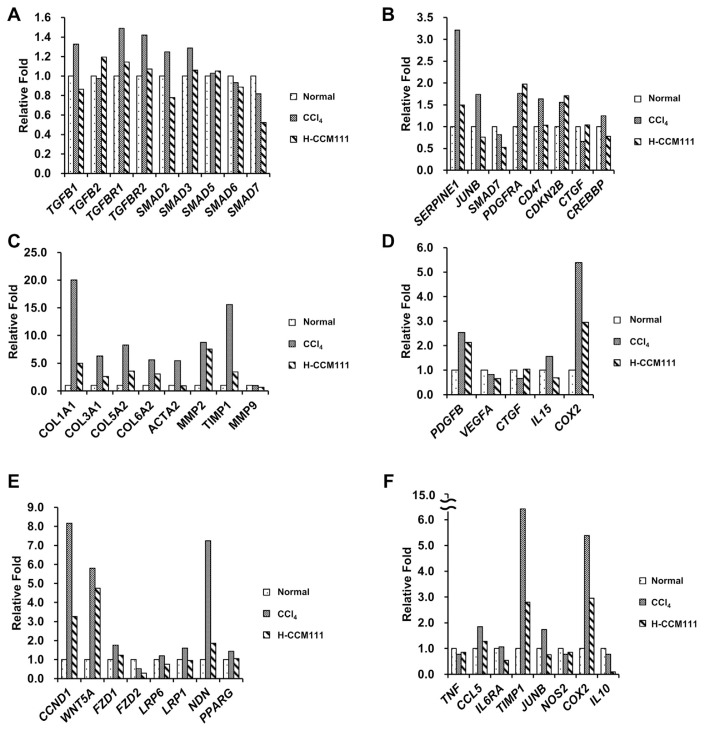
Fig. 3
Assessment of genes differentially expressed…
Fig. 3
Assessment of genes differentially expressed in liver samples. Liver samples were analyzed by…
Fig. 3 Assessment of genes differentially expressed in liver samples. Liver samples were analyzed by next-generation sequencing. The fold change was calculated by the RPKM of each group relative to the normal group. (A) TGF-β pathway genes. (B) Downstream genes of the TGF-β pathway. (C) ECM genes. (D) Cytokines. (E) Downstream genes of the Wnt pathway. (F) Downstream genes of the STAT3 pathway.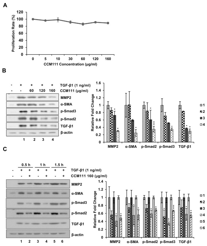
Fig. 4
CCM111 reduced TGF-β1-induced expression of…
Fig. 4
CCM111 reduced TGF-β1-induced expression of α-SMA and MMP2 and activation of the TGF-β…
Fig. 4 CCM111 reduced TGF-β1-induced expression of α-SMA and MMP2 and activation of the TGF-β signaling pathway in HSCs. (A) The cells were treated with CCM111 (0, 5, 10, 30, 60, 120 or 160 μg/ml) for 24 h. Proliferation was detected by the Alamar Blue assay. (B) The cells were starved for 24 h and treated with TGF-β1 (1 ng/ml) and CCM111 (0, 60, 120 or 160 μg/ml) for 1 h. (C) The cells were starved for 24 h and treated with TGF-β1 (1 ng/ml) and CCM111 (160 μg/ml) for 0.5, 1 and 1.5 h. Total cell lysates were prepared for western blot analysis to detect protein levels. β-actin was the loading control. The quantification data are reported as the mean ± standard deviation of three independent experiments. Student’s t-test was performed to compare the CCM111 group with the TGF-β1-only group. *P-value < 0.05, **P-value < 0.01 and ***P-value < 0.001.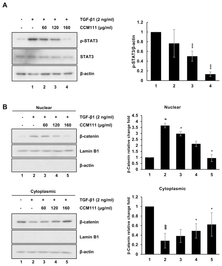
Fig. 5
CCM111 reduced TGF-β1-induced the activation…
Fig. 5
CCM111 reduced TGF-β1-induced the activation of the Wnt and STAT3 signaling pathways in…
Fig. 5 CCM111 reduced TGF-β1-induced the activation of the Wnt and STAT3 signaling pathways in HSC cells. The cells were starved for 24 h and treated with TGF-β1 (2 ng/ml) and CCM111 (0, 60, 120, 160 μg/ml) for 24 h. (A) The protein levels of p-STAT3 and STAT3 were detected. (B) The protein levels of β-catenin expression were detected. β-actin was used as the cytosol internal control, and lamin B1 was used as the nuclear internal control. The quantification data are reported as the mean standard deviation of three independent experiments. Student’s t-test compared the CCM111 treatment with the TGFβ1 only group. *P-value < 0.05, **P-value < 0.01 and ***P-value < 0.001. The control group vs. TGF-β1 only group. #P-value < 0.05, and ###P-value < 0.001.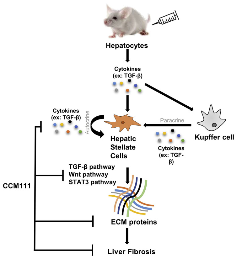
Fig. 6
CCM111 remarkably attenuated the severity…
Fig. 6
CCM111 remarkably attenuated the severity of CCl 4 -induced liver fibrosis through the…
Fig. 6 CCM111 remarkably attenuated the severity of CCl4-induced liver fibrosis through the inhibition of the TGF-β1/Smad, Wnt, and STAT3 pathways and the expression of ECM proteins and cytokines. CCM111 exhibited hepatoprotective effects in CCl4-induced experimental fibrosis, implying its potential application in clinical intervention.