Antrodia cinnamomea Oligosaccharides Suppress Lipopolysaccharide-Induced Inflammation through Promoting O-GlcNAcylation and Repressing p38/Akt Phosphorylation
Abstract
Antrodia cinnamomea (AC), an edible fungus growing in Taiwan, has various health benefits. This study was designed to examine the potential inhibitory effects of AC oligosaccharides on lipopolysaccharide (LPS)-induced inflammatory responses in vitro and in vivo. By trifluoroacetic acid degradation, two oligosaccharide products were prepared from AC polysaccharides at 90 °C (ACHO) or 25 °C (ACCO), which showed different oligosaccharide identities. Compared to ACCO, ACHO displayed better inhibitory effects on LPS-induced mRNA expression of pro-inflammatory cytokines including IL-6, IL-8, IL-1β, TNF-α and MCP-1 in macrophage cells. Further, ACHO significantly suppressed the inflammation in lung tissues of LPS-injected C57BL/6 mice. The potential anti-inflammatory molecular mechanism may be associated with the promotion of protein O-GlcNAcylation, which further skewed toward the marked suppression of p38 and Akt phosphorylation. Our results suggest that the suppressive effect of AC oligosaccharides on inflammation may be an effective approach for the prevention of inflammation-related diseases.
Figures 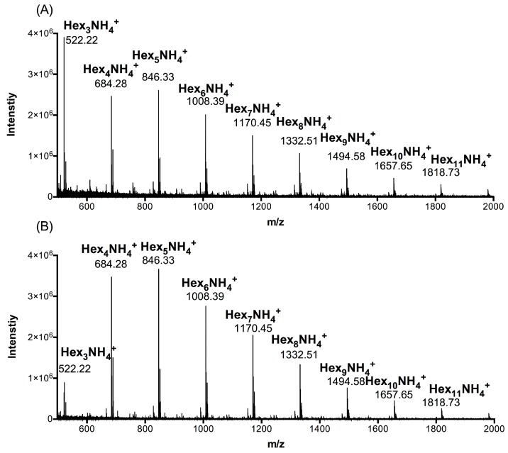
Figure 1
UPLC-MS analysis of Antrodia cinnamomea…
Figure 1
UPLC-MS analysis of Antrodia cinnamomea (AC) polysaccharides at 90 °C (ACHO) or 25…
Figure 1 UPLC-MS analysis of Antrodia cinnamomea (AC) polysaccharides at 90 °C (ACHO) or 25 °C (ACCO). (A) Polymerization degree identification of ACCO; (B) Polymerization degree identification of ACHO. Hex represents hexose.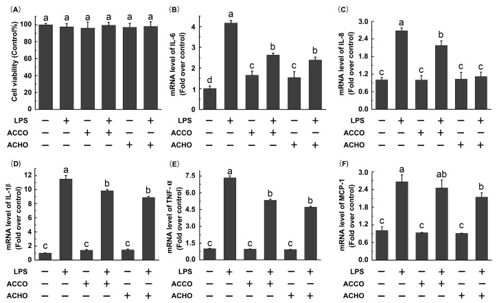
Figure 2
Effects of ACCO and ACHO…
Figure 2
Effects of ACCO and ACHO on viability and inflammatory reaction in lipopolysaccharide (LPS)-stimulated…
Figure 2 Effects of ACCO and ACHO on viability and inflammatory reaction in lipopolysaccharide (LPS)-stimulated macrophage cells. (A) RAW264.7 cells were treated with LPS (200 ng/mL), ACCO (100 µg/mL) or ACHO (100 µg/mL) for 24 h. After that, the cell viability was tested by a MTT assay; (B–F) RAW264.7 cells were pre-treated with ACCO (100 µg/mL) or ACHO (100 µg/mL) for 12 h and then exposed to LPS (200 ng/mL) for 6 h. Next, the mRNA levels of IL-6 (B), IL-8 (C), IL-1β (D), TNFα (E) and MCP-1 (F) were determined by RT-PCR analysis. Data are the mean ± SD (n = 3). Data with different superscript letters are significantly different (p < 0.05).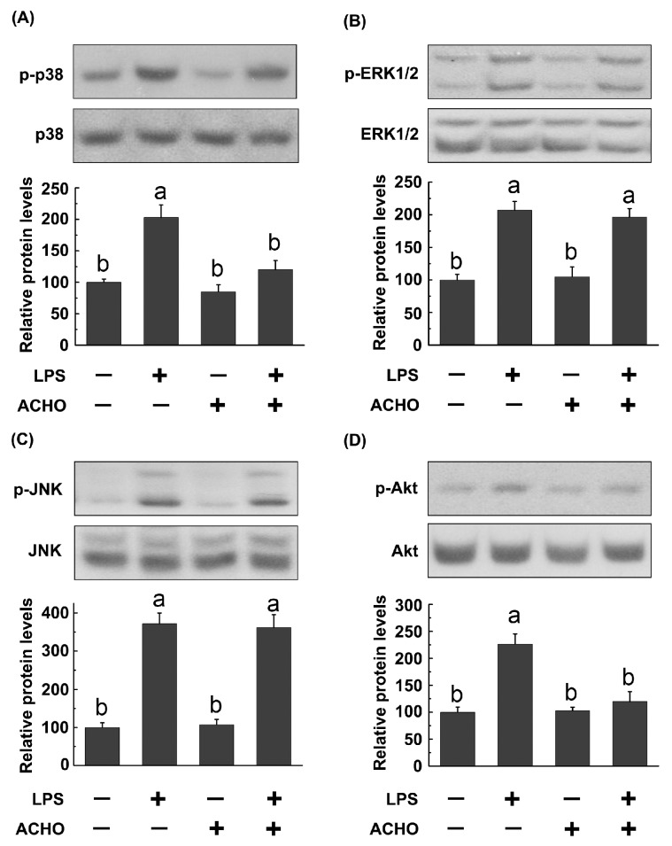
Figure 3
Inhibitory effect of ACHO on…
Figure 3
Inhibitory effect of ACHO on activation of MAPK and Akt signaling pathways in…
Figure 3 Inhibitory effect of ACHO on activation of MAPK and Akt signaling pathways in LPS-induced macrophage cells. RAW264.7 cells were pretreated with ACHO (100 µg/mL) in FBS-free medium for 12 h and then exposed to LPS (200 ng/mL) for 30 min. After that, cells were collected and the phosphorylated levels of p38 (A); ERK1/2 (B); JNK (C) and Akt (D) were determined by western blot. Data represent the mean ± SD (n = 3). Data with different superscript letters are significantly different (p < 0.05).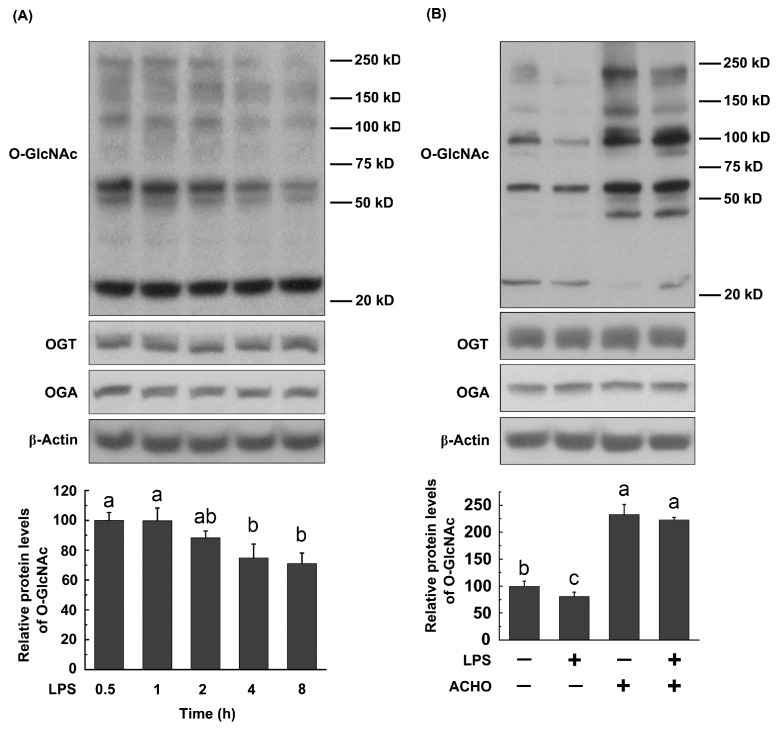
Figure 4
Effects of LPS and ACHO…
Figure 4
Effects of LPS and ACHO on protein O -GlcNAcylation in macrophage cells. (…
Figure 4 Effects of LPS and ACHO on protein O-GlcNAcylation in macrophage cells. (A) RAW264.7 cells were treated with LPS (200 ng/mL) for 0–8 h; (B) RAW264.7 cells were pretreated with ACHO (100 µg/mL) for 12 h and then exposed to LPS (200 ng/mL) for 8 h. After that, cells were collected and analyzed by western blot. Data are represented as the mean ± SD (n = 3). Data with different superscript letters are significantly different (p < 0.05).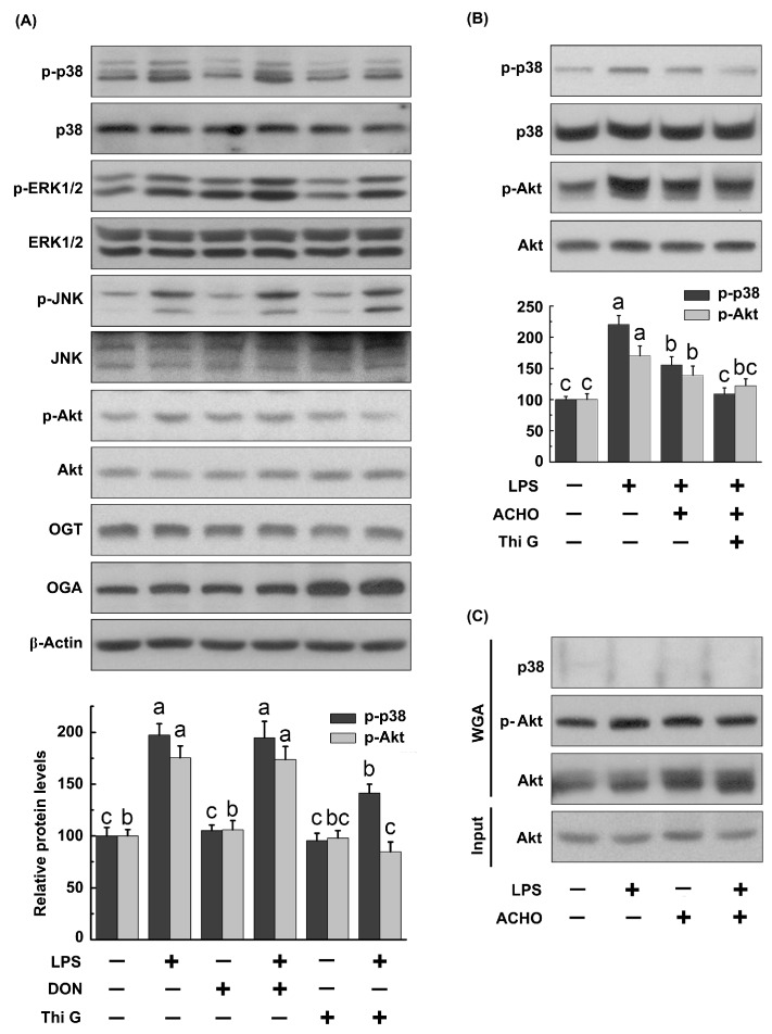
Figure 5
Suppression of MAPK and Akt…
Figure 5
Suppression of MAPK and Akt phosphorylation by protein O -GlcNAcylation in LPS-induced macrophage…
Figure 5 Suppression of MAPK and Akt phosphorylation by protein O-GlcNAcylation in LPS-induced macrophage cells. (A,B) RAW264.7 cells were pretreated with Thi G (50 nM), 6-diazo-5-oxo-l-norleucine (DON) (5 μM) or ACHO (100 μg/mL) in FBS-free medium for 12 h and then exposed to LPS (200 ng/mL) for 30 min. After that, cells were collected, and the protein samples were analyzed by western blot; (C) RAW264.7 cells were pretreated with ACHO (100 μg/mL) in FBS-free medium for 12 h and then exposed to LPS (200 ng/mL) for 30 min. Then, the O-GlcNAcylated proteins were pulled down by WGA-agarose and captured using the antibodies against p38, Akt or p-Akt. Data represent the mean ± SD (n = 3). Data with different superscript letters are significantly different (p < 0.05).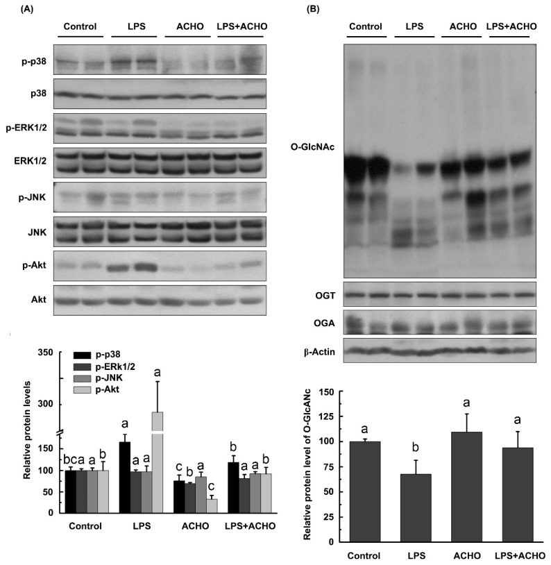
Figure 6
Effect of ACHO on MAPKs,…
Figure 6
Effect of ACHO on MAPKs, Akt and protein O -GlcNAcylation in the lung…
Figure 6 Effect of ACHO on MAPKs, Akt and protein O-GlcNAcylation in the lung tissues of LPS-injected mice. (A) Suppression of ACHO on the activation of MAPKs and Akt in lung tissues after LPS injection; (B) Reversal of ACHO on the decrease in protein O-GlcNAcylation in lung tissues after LPS injection. C57BL/6 mice were pre-treated with ACHO (1 mg/mL in drinking water) for two weeks followed by LPS injection (3 mg/kg) intraperitoneally for 24 h. After that, mice were euthanized, and lung tissues were collected for western blot assay. Data represent the mean ± SD (n = 5). Data with different superscript letters are significantly different (p < 0.05).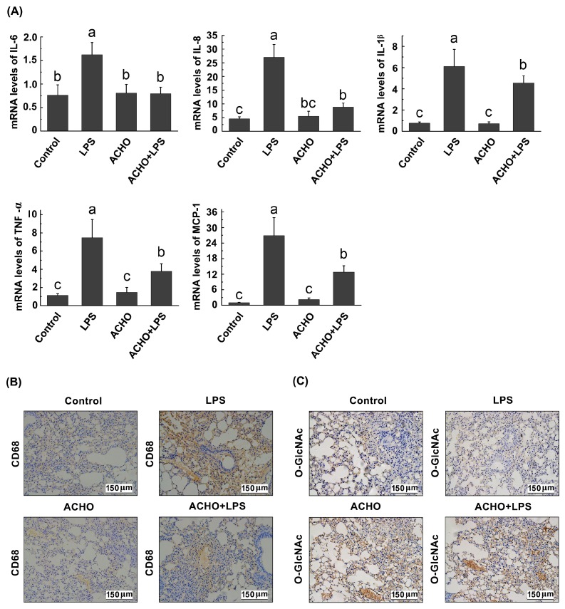
Figure 7
Effect of ACHO on transcriptional…
Figure 7
Effect of ACHO on transcriptional activation of pro-inflammatory cytokines ( A ), macrophage…
Figure 7 Effect of ACHO on transcriptional activation of pro-inflammatory cytokines (A), macrophage infiltration (B) and protein O-GlcNAcylation (C) in lung tissues of LPS-injected mice. C57BL/6 mice were pre-treated with ACHO (1 mg/mL in drinking water) for two weeks followed by LPS (3 mg/kg) injection intraperitoneally for 24 h. After that, mice were euthanized, and lung tissues were collected for RT-PCR assay or immunochemistry assay. (A) Expression of pro-inflammatory cytokines at mRNA level in lung tissues of LPS-injected mice, including IL-6, IL-8, IL-1β, TNF-α and MCP-1; (B) Effect of ACHO on CD68 (cluster of differentiation 68) production in lung tissues of LPS-injected mice (×200); (C) Effect of ACHO on protein O-GlcNAcylation in lung tissues of LPS-injected mice (×200). Data represent the mean ± SD (n = 5). Data with different superscript letters are significantly different (p < 0.05).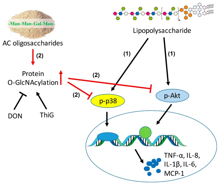
Figure 8
The schematic diagram that ACHO…
Figure 8
The schematic diagram that ACHO inhibited LPS-induced inflammation. (1) LPS induced the over-expression…
Figure 8 The schematic diagram that ACHO inhibited LPS-induced inflammation. (1) LPS induced the over-expression of IL-6, IL-8, IL-1β, TNF-α and MCP-1 at mRNA levels by up-regulation of p-p38 and p-Akt; (2) ACHO promoted the protein O-GlcNAcylation, which further suppressed phosphorylation of p38 and Akt and subsequently inhibited LPS-mediated inflammation. All figures (8)