Novel Antrodia cinnamomea Extract Reduced Cancer Stem-Like Phenotype Changes and Resensitized KRAS-Mutant Colorectal Cancer via a MicroRNA-27a Pathway
Abstract
Colorectal cancer (CRC) is one of the most common causes of death in Taiwan. Previous studies showed that Antrodia cinnamomea (AC) can treat poisoning, diarrhea, and various types of cancer. Therefore, we purified a novel ubiquinone derivative, AC009, and investigated its antitumor effects. Cell viability assays revealed that AC009 reduced the viability of several human CRC cell lines. AC009 treatment resulted in cell-cycle arrest/apoptosis, and these effects may occur via caspase and Bcl-2 signaling pathways. We demonstrated that AC009 could significantly inhibit in vivo tumor growth in xenograft mouse models. Using messenger RNA (mRNA) and microRNA (miRNA) microarrays, we found that KRAS gene expression was also regulated by AC009, possibly through specific miRNAs. AC009 also reduced cancer stem-cell marker CD44+/CD24+ expression and restored the tumor inhibition effect of cetuximab in KRAS-mutant CRC. Moreover, we found that miRNA-27a could restore the tumor inhibition effect of cetuximab in KRAS-mutant CRC cells. Taken together, our results suggest that AC009 has therapeutic potential against human wild-type and KRAS-mutant CRC.
Figures 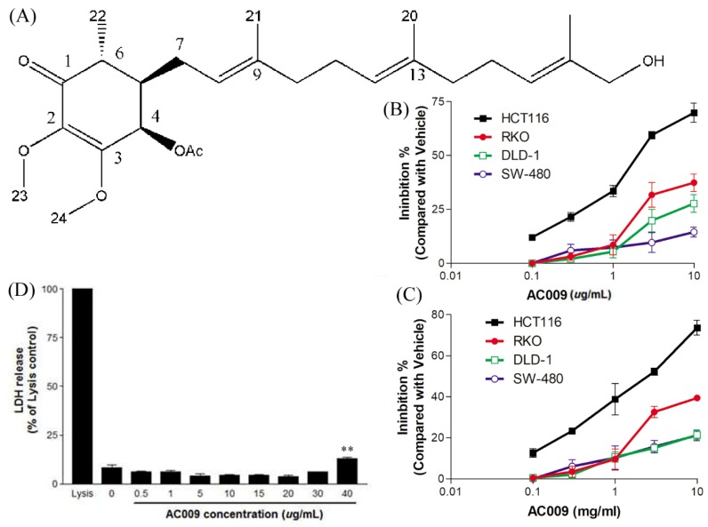
Figure 1
The structure of AC009 and…
Figure 1
The structure of AC009 and its effect on colon cancer cells viability. The…
Figure 1 The structure of AC009 and its effect on colon cancer cells viability. The structure of AC009 is shown in (A). Colorectal cancer (CRC) cells (HCT116, RKO, DLD-1, and SW-480) were starved for 48 h, cultured with DMEM (with 10% FBS) in the presence or absence AC009 (0.1, 0.3, 1, 3, 10 μg/mL). The cell viability was detected by sulforhodamine B (SRB) assay (B) and crystal violet assay (C) after 72 h; the results are presented as percentages of inhibition (n = 10). (D) The cytotoxicity of colorectal cancer cells treated with different concentrations of AC009 (HCT116 cells, 72 h treatment; n = 10, mean ± SEM, ** p < 0.01).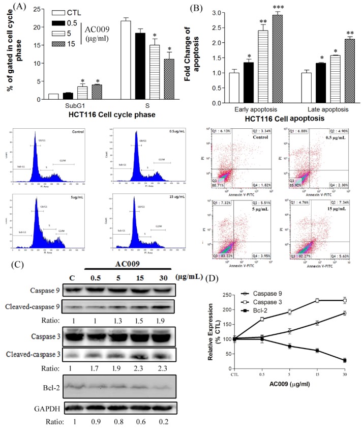
Figure 2
Effects of AC009 on HCT116…
Figure 2
Effects of AC009 on HCT116 cell-cycle arrest, the promotion of apoptosis, and caspase/Bcl-2…
Figure 2 Effects of AC009 on HCT116 cell-cycle arrest, the promotion of apoptosis, and caspase/Bcl-2 expression. A flow cytometry assay was used to detect the cell cycle and annexin V apoptosis of HCT116 cancer cells after AC009 treatment. (A) Cell-cycle change after AC009 treatment at different concentrations for 48 h. (B) Annexin V-FITC shows early- and late-phase apoptosis after AC009 treatment for 24 h. The fold-change of early and late apoptosis was analyzed (n = 10, mean ± SEM, * p < 0.05, ** p < 0.01, *** p < 0.001). (C) In caspase, the detection results show that cleaved caspase 9 and cleaved caspase 3 increased. The Bcl-2 and GAPDH expressions were also detected (n = 3). The ratios of these proteins and their controls are shown below the band. Quantitative analyses of cleaved caspase 9, cleaved caspase 3, and Bcl-2 are presented as the mean density, as determined by a densitometer (D).
Figure 3
The in vivo anti-tumor effect…
Figure 3
The in vivo anti-tumor effect of AC009. NOD/SCID mice were implanted with HCT116…
Figure 3 The in vivo anti-tumor effect of AC009. NOD/SCID mice were implanted with HCT116 CRC cells. Seven to 12 days later (tumor volume growth up to 2 mm3), AC009 was administered via intraperitoneal injection (1.5 and 6 mg/kg). (A) Tumor volume (mm3), (B) HCT116 picture of the control tumor, and after different concentrations of AC009 treatment. (C) Ratios of mice body weights (NOD/SCID mice; n = 10 per group, mean ± SEM, * p < 0.01, *** p < 0.001).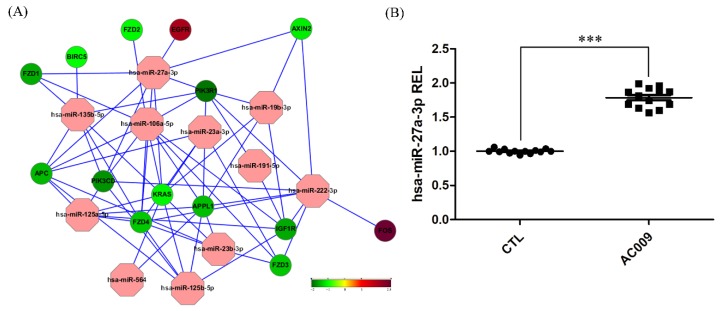
Figure 4
Microarray of microRNA (miRNA)/messenger RNA…
Figure 4
Microarray of microRNA (miRNA)/messenger RNA (mRNA) assay and qRT-PCR. ( A ) Microarray…
Figure 4 Microarray of microRNA (miRNA)/messenger RNA (mRNA) assay and qRT-PCR. (A) Microarray of miRNA assay analysis shows the up- and downregulation of specific genes after 24 h treatment with AC009. The green color indicates that mRNA expression decreased by 50%, and the red color indicates that mRNA expression increased 2.8-fold. (B) Expression levels of hsa-miR-27a-3p were normalized using the U6 small nuclear RNA (snRNA) and are expressed as REL (relative expression level); n = 13 per group, *** p < 0.001.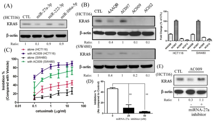
Figure 5
The effect of miRNA and…
Figure 5
The effect of miRNA and AC009 in KRAS protein expression and resensitized cetuximab…
Figure 5 The effect of miRNA and AC009 in KRAS protein expression and resensitized cetuximab antitumor ability. After treatment with miR-27a-3p, miR-222-3p, miR-106a-5p (A), AC009, or its derivatives (B), a Western blotting assay was used to detect KRAS protein expression. HCT116 or SW480 cells were grown for 24 h, starved for 48 h, and cultured with DMEM (with 10% FBS). The cell viability (SRB assay) to detect the various concentrations of cetuximab in the absence or presence of AC009 (1 μg/mL) for the HCT116 and SW480 cells is shown in (C). The effect of AC009 (1 μg/mL) on HCT116 cell viability (SRB assay) in the absence or presence of an miRNA-27a-specific inhibitor is shown in (D). The effect of KRAS protein expression influence by an miRNA-27a-specific inhibitor and AC009 is shown in (E). The ratio of KRAS and β-actin is shown below the band. All data are representative of experiments performed three times. ** p < 0.01.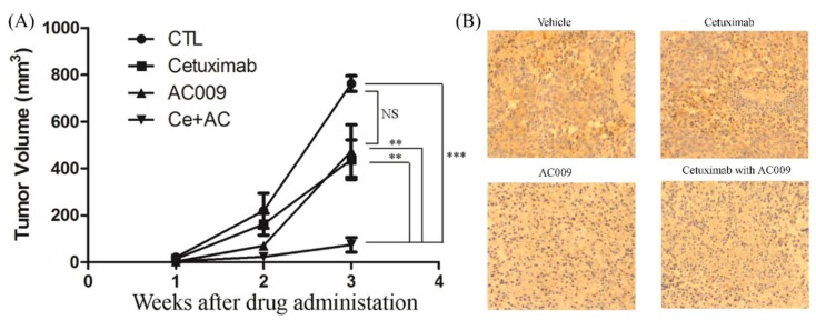
Figure 6
The effect of AC009 resensitizes…
Figure 6
The effect of AC009 resensitizes the cetuximab anti-tumor ability in in vivo models.…
Figure 6 The effect of AC009 resensitizes the cetuximab anti-tumor ability in in vivo models. The tumor volume (mm3) of cetuximab in the absence or presence of AC009 inhibited HCT116 tumor growth in the NOD/SCID mouse xenograft (A). The expression of KRAS in a HCT116 tumor xenograft was studied (B). ** p < 0.01, *** p < 0.001.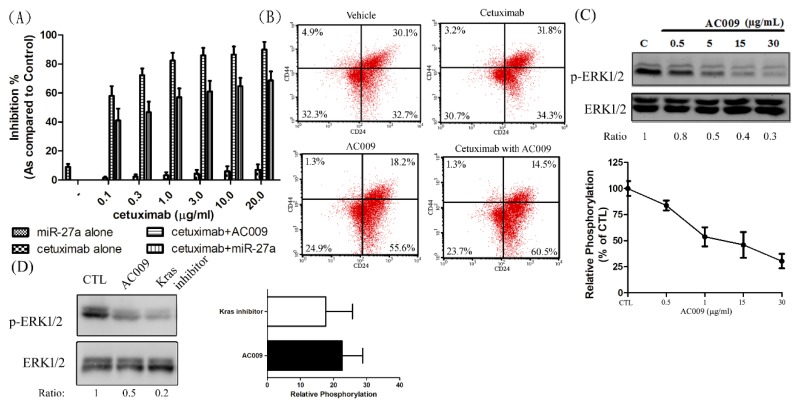
Figure 7
The effect of miR-27a on…
Figure 7
The effect of miR-27a on the resensitized cetuximab anti-tumor ability, and the effect…
Figure 7 The effect of miR-27a on the resensitized cetuximab anti-tumor ability, and the effect of AC009 in cancer stem-cell marker expression. HCT116 cells were transfected with miR-27a mimics and grown for 24 h, starved for 48 h, cultured with DMEM (with 10% FBS), and treated with various concentrations of cetuximab in the absence or presence of AC009 (1 μg/mL), and cell viability was detected (SRB assay) (A). A flow cytometry assay was used to detect CD44 and CD24 expression on the HCT116 cell surface (B). After AC009 treatment (C) or KRAS inhibitor treatment (D), the ERK phosphorylation was studied by Western blotting, and the mean density was determined by a densitometer. The ratio of p-ERK1/2 and ERK1/2 is shown below the band. All data are representative of experiments performed three times.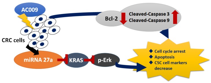
Figure 8
The therapeutic effects and mechanisms…
Figure 8
The therapeutic effects and mechanisms of AC009 in colorectal cancer (CRC) cell treatment.…
Figure 8 The therapeutic effects and mechanisms of AC009 in colorectal cancer (CRC) cell treatment. After Antrodia cinnamomea (AC) extract AC009 treatment, CRC cells may undergo apoptosis through the caspase-3- and caspase-9-dependent signaling pathways and further downregulate the Bcl-2 expression. In addition, AC009 also caused cell-cycle arrest in CRC cell treatment. AC009 also promoted miRNA-27a expression and further inhibition of KRAS expression. Then, p-Erk, which is a downstream protein of KRAS signaling pathway, was also downregulated and further caused the cancer stem cell (CSC) markers to decrease after AC009 treatment in CRC. All figures (8)